Human beings have all the time been fascinated by the brain.
Although scientific knowledge about this 1.3 kg of fragile substance embedded in our cranium has long been incomplete, dazzling technical breakthroughs made lately at the moment are ushering in a Golden Age of molecular neuroscience.
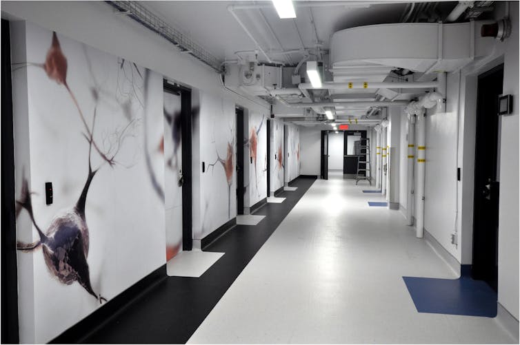
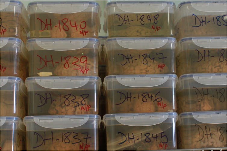
These breakthroughs have been made possible partly due to brain banks, which preserve human brains in the perfect possible conditions for scientific research. Here in Montréal, we’ve one among the world’s largest such banks, the Douglas-Bell Canada Brain Bank (DBCBB), founded in 1980 on the Douglas Hospital.
The DBCBB, which receives several brains every month, has collected over 3,600 specimens thus far. Every 12 months, its team processes dozens of tissue requests from scientists in Québec, Canada and abroad, preparing some 2,000 samples for research.
Over the past 40 years, these efforts have led to a substantial variety of discoveries about different neurological and psychiatric diseases.
As a full professor within the department of psychiatry at McGill University, researcher on the Douglas Research Centre and director of the DBCBB since 2007, I work in close collaboration with Dr. Gustavo Tureckico-director of the DBCBB and accountable for the component dedicated to psychiatric illnesses and suicide.
(Naguib Mechawar), Provided by the creator
A transient history of research on the human brain
Scientists only began to discover the microscopic elements that make up the human brain within the second half of the nineteenth century.
That was when brains were preserved for the primary time in formalin, an answer that preserves biological tissue in order that it could actually be handled more easily and stored over a long term.
At the identical time, precision instruments and protocols were being developed that made it possible to look at the microscopic characteristics of nervous tissue.
Until the center of the twentieth century, researchers were mainly satisfied with preserving the brains of patients, taken during autopsies, so that they could use them to discover possible macroscopic or microscopic changes linked to either neurological or psychiatric symptoms.
This is in reality what the German neurologist Alois Alzheimer did when he analyzed the brain of one among his patients affected by dementia. In 1906, he described, for the primary time, the microscopic lesions which characterize the disease that now bears his name.
Until the tip of the Nineteen Seventies, quite a few collections of brain specimens preserved in formalin were inbuilt hospital environments, a bit like the cupboards of curiosities of olden days.
Towards the tip of the twentieth century, latest experimental approaches were developed allowing the high-resolution evaluation of cells and molecules inside biological tissues.
It then became needed to gather and preserve human brains, obtained with the consent of the person or his or her family, in conditions compatible with modern scientific techniques.
Researchers began freezing one among the cerebral hemispheres as a way to measure its various molecular components. The other hemisphere was preserved in formalin for use for macroscopic and microscopic anatomical studies.
This was the context by which the Douglas-Bell Canada Brain Bank was created.

(Naguib Mechawar), Provided by the creator
New experimental approaches are yielding results
Leading researchers from many universities around the globe now use DBCBB samples to advance their research. This, in fact, includes numerous teams in Québec.
For example, along with his team from the Douglas Research Centre, which is affiliated with McGill University, Jude Poirier discovered that the APOE4 gene is a risk factor for Alzheimer’s disease. More recently, the team of Gilbert Bernierprofessor within the department of neuroscience at Université de Montréal, discovered that the lesions characteristic of this disease are related to abnormal expression of the BMI1 gene.
With regard to psychiatric illnesses, and more specifically depression, major progress has been made recently by the McGill Group for Suicide Studies.
Using cutting-edge methods to isolate and analyze human brain cells, Turecki’s team has succeeded in exactly identifying the cell types whose function is affected in men who’ve suffered from major depressionafter which discovering that the cell types involved on this illness differ between men and girls.
These experimental approaches generate huge data sets that will be examined in subsequent studies. This is the case, for instance, of labor carried out in my laboratory, which identified signs of persistent changes in neuroplasticity throughout the prefrontal cortex of individuals with a history of child abuse. In fact, the studies mentioned above enabled us to find at the very least one among the cell types involved on this phenomenon.
In short, the experimental methods we’ve today allow us to interrupt the brain down into its elementary components as a way to understand its functions and dysfunctions.

(Naguib Mechawar), Provided by the creator
Identify, prevent, screen and treat
Thanks to the exertions and dedication of the whole DBCBB team, in addition to the unfailing support of all its partners, patrons (often anonymous) and funding bodies — particularly the FRQS research fund and Québec’s suicide research network, the Quebec network on suicide, mood disorders and associated disorders — this invaluable resource has not only managed to survive, but to grow and grow to be one among the biggest brain banks on this planet.
There is every reason to imagine that, within the years to come back, the DBCBB will play a vital role within the increasingly precise identification of the biological causes of brain diseases, and, in consequence, will contribute to the identification of latest targets for higher approaches to prevention, screening and treatment.


